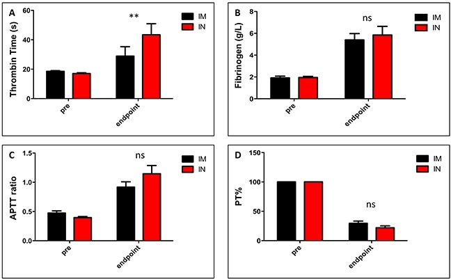
Figure 3. Markers of coagulation in SUDV-infected ferrets.

Ferrets were infected with SUDV intramuscularly (shown in black) or intranasally (shown in red). Markers of coagulation in the blood were analyzed using the STart4 analyzer. For each ferret, blood samples were taken pre-infection and also at time of death (days 7-9). Plasma samples were mixed with a known amount of thrombin and the time until clotted was measured (A). A normal value = 21s while higher values indicate the presence of coagulation factors. Plasma clotting times were assayed and fibrinogen levels were calculated based on a standard curve (B). Activated partial thromboplastin times (C) and % partial thromboplastin times (D) were measured from plasma before infection and at time of death.
