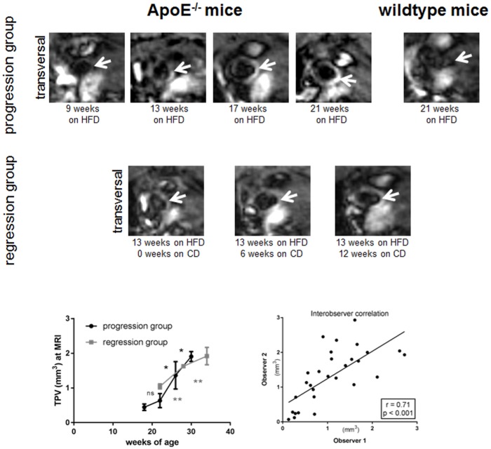
Fig 4. Progression and regression of atherosclerosis at 7T MRI.
MRI in vivo showed a transversal slice orientation through the ascending aorta. Progression of atherosclerotic lesion (arrow) in ApoE-/- mice can be visualized over time. Calculated TPV showed a more linear increase in case of mouse group reswitching to chow diet (CD) 13 weeks after starting HFD. Interobserver analysis showed a strong and significant correlation of r = 0.71; p<0.001. No plaque was detectable in wildtype mice. (n.s.—non significant, *p<0.05, **p<0.01).

