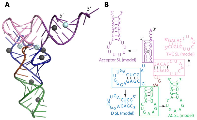
Figure 1.
Structures of FL yeast tRNAphe and its model helical fragments (HF) with their predicted folds. (A) Three-dimensional folded structure of yeast FL tRNAphe (PDB: 1ehz) and (B) FL tRNA secondary structure and the model HF derived from each stem. In panel (A) Mg2+ (cyan) and Mn2+ (black) ions associated with tRNA are shown as spheres. The colors of each structural element are the same in both panels. FL tRNA tertiary contacts are provided in Figure S1. The sequences of the model HF are the same as in FL tRNAphe and are predicted to form the shown hairpins by the Mfold server for each model sequence.

