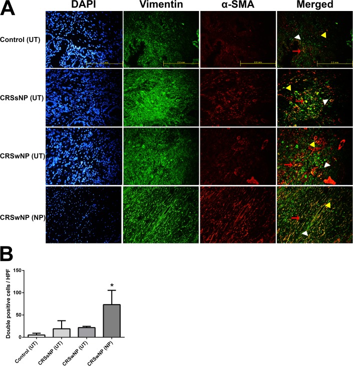
Fig 1. Double–immunofluorescent staining of myofibroblasts in the tissues of the four groups.
Double-immunofluorescence staining was undertaken to colocalize the cells with Vimentin (green color)/ α-SMA (red color) among the groups (A). The number of double positive cells (Vimentin+ α-SMA+) was considerably higher in the NP tissues of the CRSwNP group compared to the other groups (*p < 0.05) (B). Vimentin: white arrow head, α-SMA: yellow arrow head, double positive cells: red arrow. Triple tests were performed on all of the experiments.

