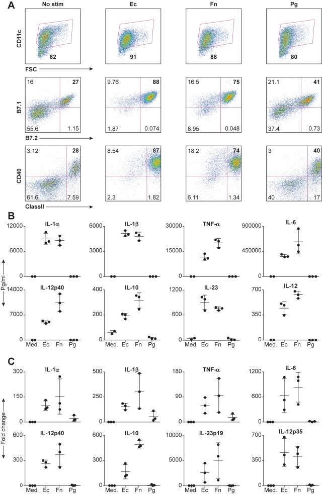
Fig 1. Inflammatory cytokine production from DCs is extinguished in the presence of P. gingivalis.
(A) DCs from B10.A-Rag2-/- mice were either unstimulated (medium) or stimulated on the sixth day with 5x10⁷ bacteria in a 24-well plate for 16h at 37°C. Cells were harvested, stained with directly labeled monoclonal antibodies and analyzed by FACS analysis. Cells were gated for the CD11c positive population and analyzed for the costimulatory molecules B7.1, B7.2 and CD40, and the antigen-presenting molecule, MHC class II. Numbers indicate the percentage of the population in the gate or quadrant (B) Same as (A) Culture CSN were analyzed for the presence of various cytokines using Searchlight protein arrays. (C) Same as (B) except the co-culture period was 6h, at which time we isolated total RNA using the RNeasy kit and analyzed it by Real-time RT-PCR. The data are expressed as the mean ± SD of three independent experiments.

