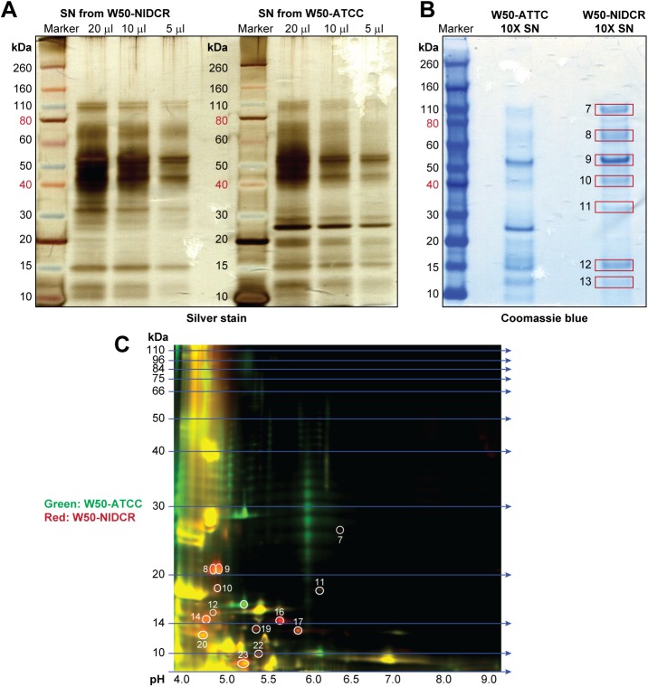
Fig 4. Comparative proteomic of particle-free SN from W50-NIDCR and W50-ATCC.
(A) Various amounts of particle-free SN from W50-ATCC and W50-NIDCR were resolved by one-dimensional SDS-PAGE and protein bands were visualized by Silver staining. (B) Coomassie staining of the same SNs that had been concentrated to 10X as described indicated in the materials and methods. (C) Same as (A) except particle-free bacterial SN from W50-ATCC and W50-NIDCR were labeled green or red, respectively, and resolved by two-dimensional difference in-gel electrophoresis.

