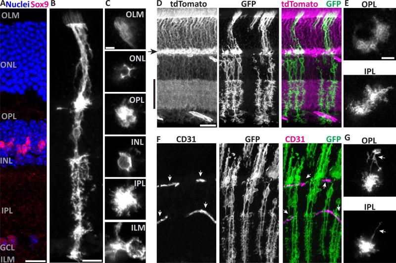
Figure 1. Müller cells have radially-oriented bipolar stalks with layer-specific branching morphology.
A) Cross-section of adult mouse retina stained with Sox9 to show Müller glia (MG) nuclei in INL, and counterstained with nuclear marker (Hoechst) to reveal major retinal layers. Text labels indicate cell body layers (GCL, INL, ONL), synaptic layers (IPL, OPL), and approximate location of limiting membranes (ILM, OLM). B,C) Morphology of individual mGFP-expressing MG from GLAST-CreER; mTmG mice, viewed in cross-section (B) or en face (C). C depicts the same cell imaged at different planes of a flat mount. Image in B is scaled to approximately match layers in A. Note morphological specializations at each layer: OLM, microvilli; ONL, processes intercalated between photoreceptor cell bodies; OPL and IPL, extensive fine branches; INL, MG cell soma; ILM, broad branches and endfeet. D,E) Retinas with dense MG labeling, showing confluence of MG arbors in synaptic layers and limiting membranes. D: Cross-section view; tdTomato fluorescence from unrecombined mTmG cells (left) counterstains synaptic layers (arrowhead, OPL; vertical bar, IPL). E: Flat mount en face view, showing confluent arbors of neighboring MG. F,G) MG branches are closely associated with CD31+ blood vessels (F, arrows). MG often have long horizontal branches (G, arrows) in the plexiform layers which can terminate on blood vessels. Scale bars (in µm): 15 (A,B), 5 (C), 25 (D,F), 10 (E,G).

