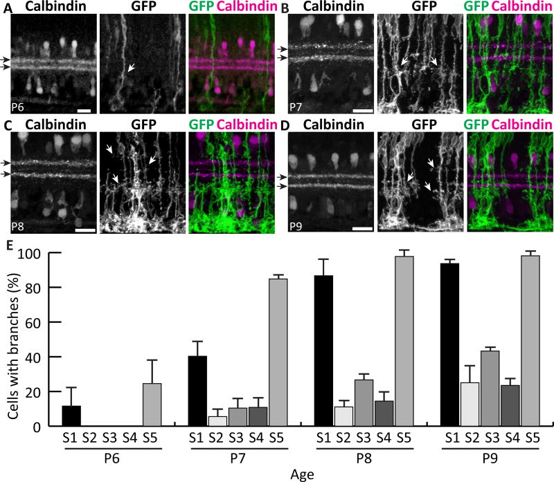
Figure 8. Müller cells first branch at stereotyped depths within the IPL.
Retinal cross sections of different developmental ages double-stained for mGFP-labeled MG and Calbindin to mark S2 and S4 (black arrows). A) At P6, most cells are unbranched, but occasional MG branches (white arrows) are seen in S1 and S5. B) At P7, branches in S1 and S5 become more abundant and the first branches in central IPL emerge. C,D) Between P8 (C) and P9 (D), branching in all layers continues to increase. Most cells branch in S1 and S5 by P9, but central layer branches continue to emerge after this time. E) Percentage of cells per retina that have branches in each IPL sublamina, measured at P6–9. Scale bars: 20 µm.

