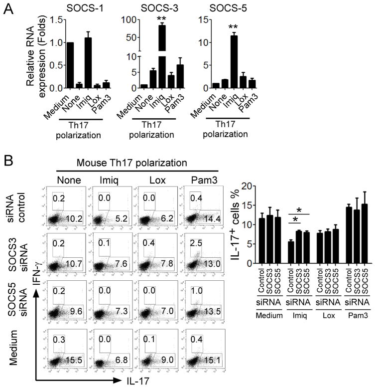
Figure 6. Induction of SOCS3 and SOCS5 is responsible for the STAT3 signaling inhibition and Th17 differentiation mediated by TLR7.
(A) Imiq but not Lox treatment significantly promoted SOCS3 and SOCS5 expression in CD4+ T cells in the presence of Th17 polarization condition. Mouse naïve CD4+ T cells were cultured in Th17 differentiation medium for 6 days in the presence or absence of Imiq, Lox or Pam3. mRNA expression levels of SOCS-1, SOCS3 and SOCS5 were determined by real-time PCR. Relative mRNA expression level of each gene was normalized to GAPDH expression and then adjusted to the level in naïve CD4+ T cells. Data shown are mean ± SD from triplicate experiments, and paired t-test was performed. ** p<0.01, compared with Th17 polarization medium only group. (B) Knockdown of SOCS3 and SOCS5 genes in CD4+ T cells dramatically alleviated the imiquimod-mediated suppression of Th17 development. Mouse CD4+ T cells were transfected with specific siRNAs against SOCS3 or SOCS5, as well as related scrambled siRNA controls, and then cultured in Th17 polarization medium in the presence or absence indicated TLR ligands for 6 days. IL-17- and IFN-γ-producing cells were evaluated using flow cytometry analysis after stimulation with PMA and ionomycin for 5 hours. Results shown in the right panel are the summary of IL-17-producing cells in differentiated Th17 cells with indicated TLR ligand and siRNA treatments obtained from three different individual experiments. Data shown are mean ± SD, and *p<0.05, compared with the scrambled siRNA control group determined by paired t-test.

