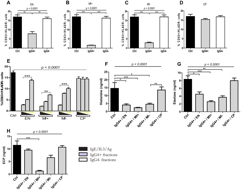
Fig 5. Depletion of IgG4 abrogates the suppressive capacity of IgG positive fractions from plasma of LF infected individuals.
Freshly isolated granulocytes from healthy blood donors (n = 9) were stimulated with IL-3 (2 ng/ml), anti-IgE (25 ng/ml) and Brugia antigen extracts (10 μg/ml) alone (dark bars) or in presence of 2.5 μg/ml of IgG4 negative fractions obtained from IgG enriched fractions (n = 8) (light bars) from EN (A), Mf+ (B), Mf- (C), CP (D) or the corresponding IgG4 positive fractions (n = 8) (grey bars) and increasing concentration (1.25 μg/ml, 2.5 μg/ml and 5 μg/ml) of IgG4 positive fractions from different groups (E). Cells were stained with anti-CD63 and HLADR antibodies. Activated granulocytes were characterized as CD63+/HLADR- cells. The release of histamine (F), neutrophil elastase (G) and eosinophil cationic protein (H) in supernatants from cultures with IgG4 positive fractions was assessed after 18 hours. Bars represent means ± SEM. Graphs are representative of 3 independent experiments. Statistical comparison was based on Kruskal-Wallis one-way ANOVA followed by Dunn post-hoc test. The indicated p-values refer to the significance level among all groups according to Kruskal-Wallis test. Asterisks indicate the level of differences after Dunn’s multiple comparisons test;*: p<0.05; **: p<0.01; ***: p<0.001.

