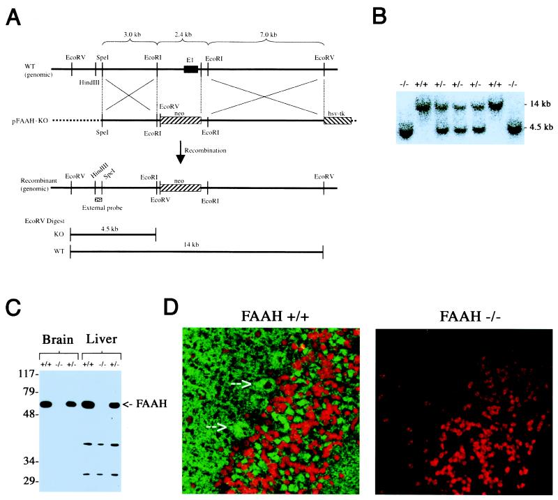
Figure 1.
Generation and biochemical characterization of FAAH−/− mice. (A) The genomic structure surrounding the deleted FAAH exon 1 (E1). Only relevant restriction sites are designated. The deleted E1 exon encodes amino acids 1–65 of the FAAH protein. (B) Southern blot analysis of EcoRV-digested genomic DNA by using the indicated probe (External probe in A), where 4.5- and 14-kb bands correspond to FAAH−/− and FAAH+/+ genotypes, respectively. (C) Western blot analysis of tissues from FAAH+/+, FAAH+/−, and FAAH−/− mice demonstrating the selective absence of FAAH protein in FAAH−/− animals. (D) Confocal microscopy immunofluorescence images of cerebellar sections of FAAH+/+ (Left) and FAAH−/− (Right) mice. Green signal, anti-FAAH; red signal, propidium iodide (stains nuclei). Arrowheads highlight intense FAAH immunoreactivity in the cell bodies of Purkinje neurons (Left).

