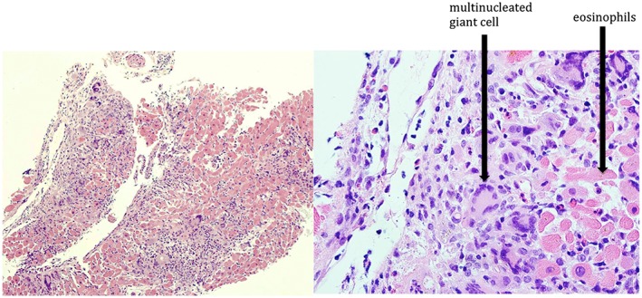
Figure 4.

Endomyocardial biopsy demonstrating inflammatory infiltration of cardiac myocardium with a haematoxylin and eosin stain (left). Magnified view (right) demonstrating numerous giant cells within inflammatory infiltrate consisting of lymphocytes, histiocytes, and eosinophils.
