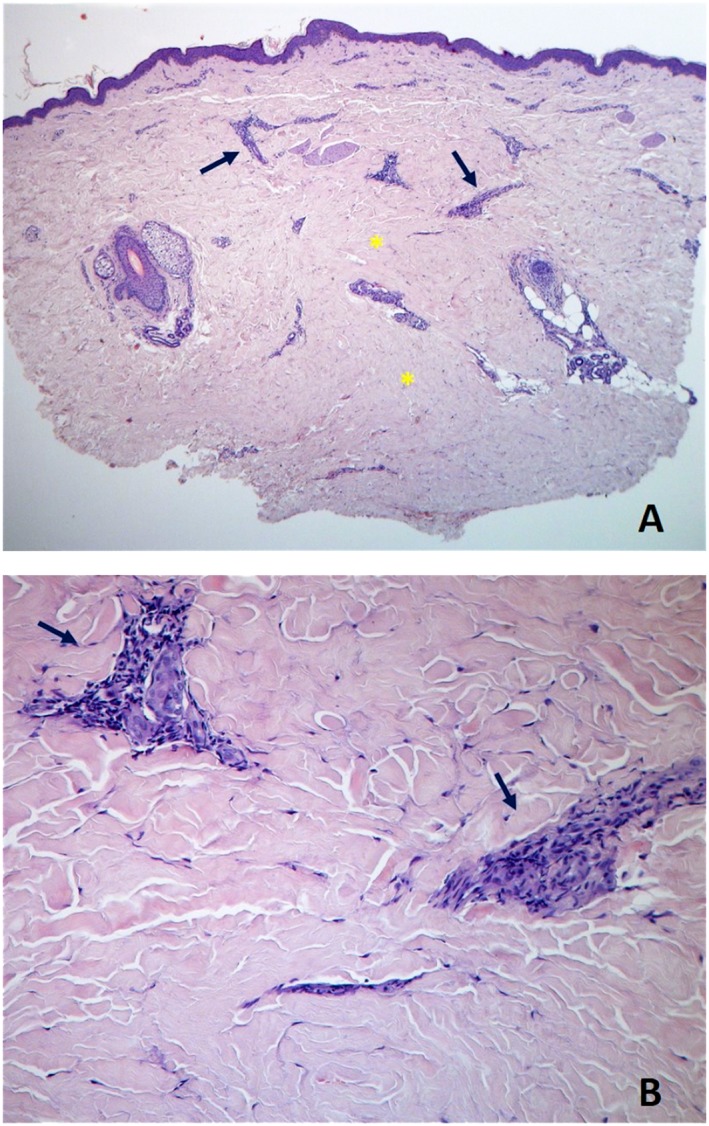
Figure 1.

Skin biopsy specimen from right forearm demonstrates dense sclerotic collagen in the reticular and deep dermis (asterisks), sparse lymphocytic infiltrate (arrows), and relative preservation of some cutaneous annexes, compatible with early skin involvement [haematoxylin‐eosin, ×40 panel (A), ×200 panel (B)].
