Figure 2.

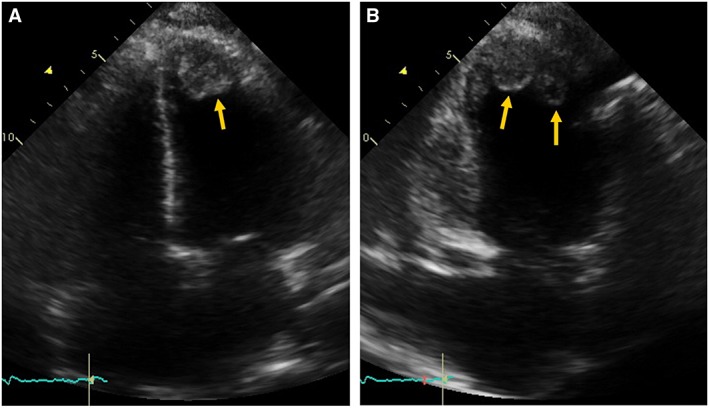
Transthoracic echocardiography. Apical four chambers view (A) and two chambers view (B) showing dilated cardiac chambers and two apical left ventricular thrombus (arrows).

Transthoracic echocardiography. Apical four chambers view (A) and two chambers view (B) showing dilated cardiac chambers and two apical left ventricular thrombus (arrows).