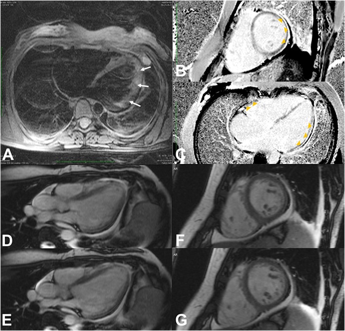
Figure 3.

Cardiac Magnetic Resonance. Patchy intramural hyperintensity of the lateral wall of the left ventricular on axial T2‐weighted image [arrows, (A)] suggests myocardial oedema. Inversion‐recovery sequences after intravascular gadolinium administration [short axis and long axis four chambers view, (B) and (C), respectively] show subepicardial delayed enhancement (arrowheads) of the inferior, infero‐lateral and lateral walls of the left ventricular, free wall and septal myocardial of the right ventricular, and pericardium. Steady‐state free precession sequences [long axis three chambers view (D–E), and short axis (F–G) during systole (D and F), and during diastole (E and G)] show pericardial effusion, as well as left sided pleural effusion. Moving images (Movies S1 and S2, Supporting Information) reveal diffuse hypokinesia of both ventricles (more severe at the inferior and infero‐lateral walls of the left ventricle).
