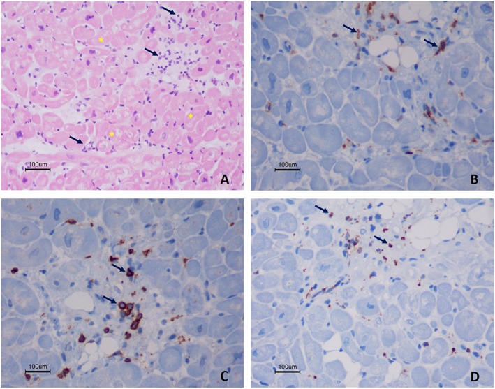
Figure 4.

Histology and immunohistochemistry findings from right ventricular endomyocardial biopsy, of active autoimmune myocarditis. Haematoxylin and eosin staining (×200) shows inflammatory infiltrates (arrows) associated with necrosis of adjacent myocytes (cytoplasmic vacuolation and nuclear atypia of myocytes—asterisks) (A). The immunohistochemistry (original magnification of 400×) shows the presence of CD4 positive cells (B), CD8 positive cells (C), and scattered macrophages [CD68 positive cells, (D)]. There is immunostaining for C4d.
