. 2017 Aug 3;6:e27711. doi: 10.7554/eLife.27711

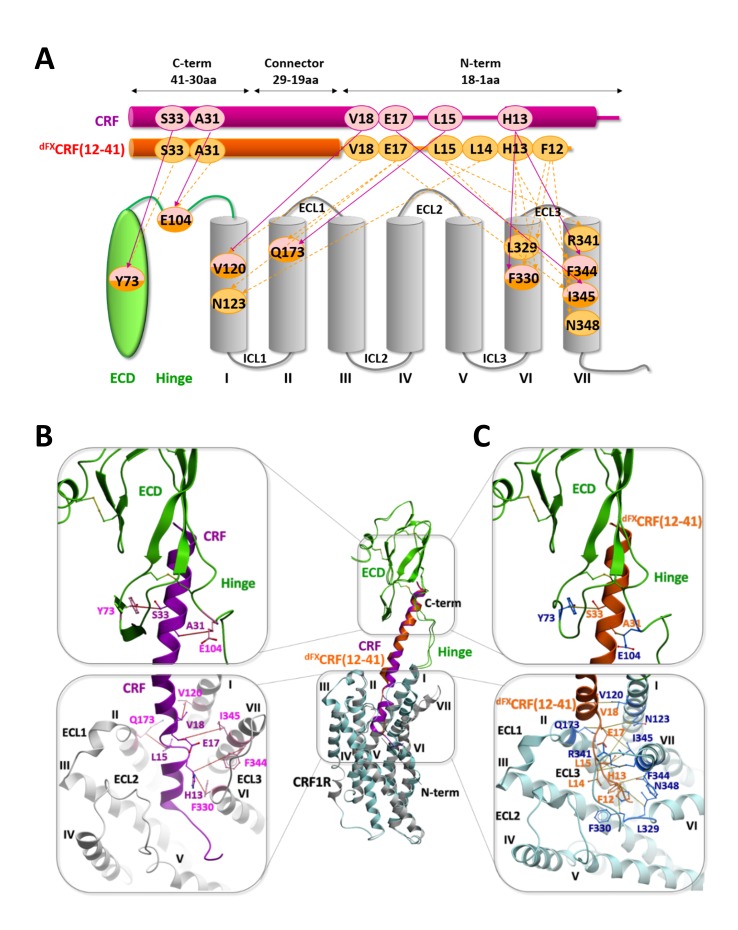
Figure 5. Comparison of the agonist-bound model CRF-CRF1R with the antagonist-bound model dFXCRF(12-41)-CRF1R.

(A) Pair-wise crosslinking hits of CRF (magenta) and dFXCRF(12-41) (orange). (B, C) Overall view and zoom into the details of the superimposed models of the CRF (magenta)-CRF1R (grey) complex and the dFXCRF(12-41) (orange)-CRF1R (light blue) complex. The framed sections are magnified in the panels (B) (CRF-CRF1R) and (C) (dFXCRF(12-41)-CRF1R). Residue pairs shown in sticks and connected by dotted lines (distances are presented in Table 2) indicate distance restraints derived from pair-wise crosslinking.

DOI: http://dx.doi.org/10.7554/eLife.27711.013

Figure 5.

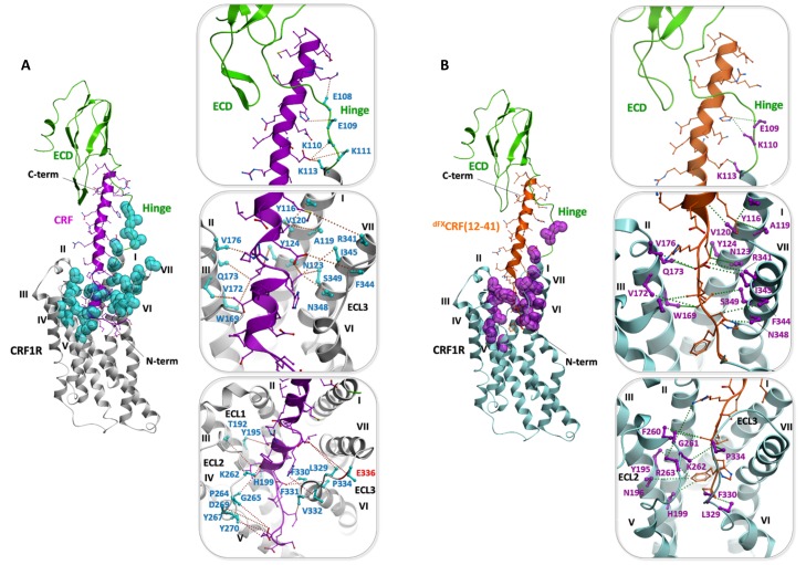
Figure 5—figure supplement 1. Validation of the peptide-CRF1R complexes by photo-crosslinking hits.

Figure 5—figure supplement 1.

(A) Validation of the CRF-CRF1R complex. (B) Validation of the dFXCRF(12-41)-CRF1R complex. An overview of each model is shown on the left. The peptide binding pocket is magnified in the panels on the right, showing the side view of the hinge region (top), helices I, II and VII (middle) and helices III, V and VI (bottom). The dotted lines connect positions in CRF1R identified by Azi photo-crosslinking screen (cyan or magenta, sticks are shown only up to Cβ) with the nearest non-hydrogen atom of CRF (magenta) or dFXCRF(12-41) (orange). Peptide side chains are shown as sticks. All distances shown, except for residue E336ECL3 (red label), are shorter than 9 Å.
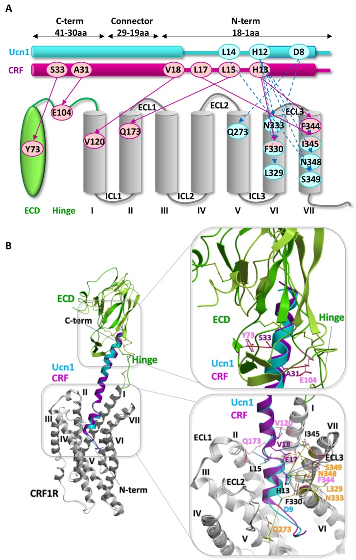
Figure 5—figure supplement 2. Comparison of the two agonist-bound models Ucn1-CRF1R and CRF-CRF1R.

Figure 5—figure supplement 2.

(A) Pair-wise crosslinking hits of Ucn1 (cyan) and CRF (magenta) with CRF1R. Notably, pair-wise crosslinking was performed using different electrophilic moieties and a different experimental approach in the two cases. For the Ucn1-CRF1R complex, the electrophilic p-2′-fluoroacetylphenylalanine (Ffact) was genetically incorporated into the receptor and Cys residues were incorporated into the ligand. For the CRF-CRF1R complex, the Lys(ClAc) electrophilic moiety was incorporated into the ligand and Cys in the receptor. The two different approaches yielded two distinct sets of pair-wise crosslinking hits, sharing only one overlapping restraint to F3306.56 (H13CRF, H13Ucn1). The models of both peptide complexes, however, are absolutely consistent with each other, validating the predictive power of the models. (B) Side view of the superimposed models of the Ucn1 (cyan)-CRF1R complex and the CRF (magenta)-CRF1R complex. The peptide-binding pocket is magnified showing the interactions with ECD and TMD. Residue pairs shown in sticks (Ucn1: cyan and yellow, CRF: magenta and pink) and connected by dotted lines (Ucn1: blue, CRF: red, distances are presented in Table 3) indicate distance restraints derived from pair-wise crosslinking.