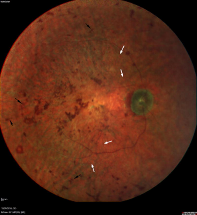
Fig 1.
Multicolor 55 Degree Image of the retina (right) reveals a pallorous optic nerve with severely attenuated retinal arterioles (white arrows). Central atrophy is evident with associated coarse, placoid pigmentation in the macula. The midperiphery reveals diffuse retinal pigment epithelial atrophy with associated bone spicule-like pigmentation (black arrows). Similar findings were present in the left eye.

