Figure 2.

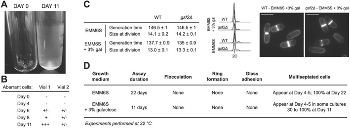
Addition of galactose and deletion of the flocculin‐encoding gene gsf2. (A) S. pombe cells were grown in the ministats as in Figure 1 in EMM6S supplemented with 3% galactose at 32°C. While this prevented flocculation, a ring of healthy cells accumulated at the top of the liquid column, introducing a strong bias for conducting any selection experiments in these devices (data not shown, similar to Figure 1D). In addition, we observed the formation of a thin film of cells adhering to the glass wall of the vessels, covering the entire surface in contact with the cell culture. A clean empty tube (day 0) is shown for comparison. The tube at day 11 was emptied, highlighting the presence of cells on the wall of the glass vessel. (B) The presence of galactose in these experiments did not prevent the random accumulation of aberrant cells in the device. The results for two independent cultures are shown. The scores were assigned as in Table 1. (C) Deletion of the gsf2 flocculin‐encoding gene has no visible effect on cells exponentially growing in batch cultures in EMM6S or EMM6S + 3% galactose at 32°C. Left panel: generation time and size at division for the indicated strains and growth conditions. WT, Wild type. Standard errors of three independent experiments are shown. For cell size at division, n > 50 for each independent experiment. Middle panel: DNA content analysis. Right panel: blankophor staining for cultures in EMM6S + 3% galactose. Scale bar = 10 μm. (D) Characterization of ministat assays using gsf2Δ cells grown in the indicated media at 32°C.
