Fig. 6.
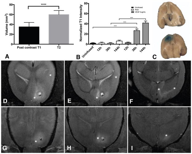
Volume of BBB breakdown is less than vasogenic edema. The volume of postcontrast T1 hyperintensity is significantly lower than the volume of T2 hyperintensity at 144 hours (A). Normalized T1 values in the juxtacanalicular region are elevated at 36 hours and beyond with VEGF infusion (B). Terminal Evans blue infusion (C) confirmed that the region of BBB breakdown was limited to the juxtacanalicular region in animals receiving VEGF (lower). Evans blue infusion (C) in animals receiving control solution (upper) did not reveal BBB breakdown. Note the expected coloration in choroid plexus bilaterally. Representative axial MR images from 1 animal demonstrate that the vasogenic edema (asterisks) on T2-weighted images extends beyond the juxtacanalicular regions (D–F). Postcontrast T1 enhancement (asterisks) in the same animal is limited to the juxtacanalicular region (G–I). ****p < 00001. Figure is available in color online only.

