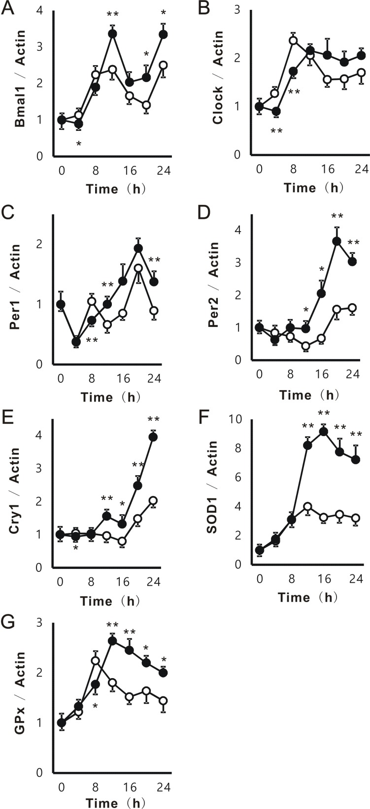
Figure 2.
Effects of PFE on circadian expression of clock genes in NIH3T3. After the treatment with serum-rich medium, NIH3T3 cells were cultured with or without PFE at 100 μg/mL for 0, 4, 8, 12, 16, 20, and 24 h. Cells were collected at each time, and the mRNA expression of Bmal1 (A), Clock (B), Per1 (C), Per2 (D), Cry1 (E), SOD1 (F), and GPx (G) in NIH3T3 cells was evaluated by real-time RT-PCR. The mRNA expression of β-actin was used to correct the expression of each mRNA. Each point (●; PFE or O; control) represents the mean and SE (n=4). Asterisks denote significant differences from the control at *p<0.05 and **p<0.01.

