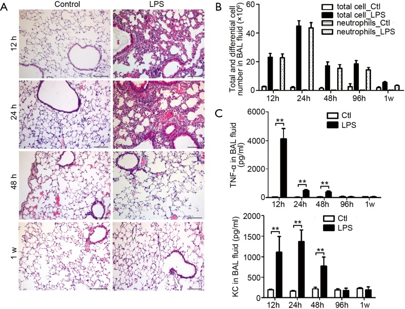
Figure 3.
Lung inflammation in mice received intranasal LPS inhalation. (A) Hematoxylin and eosin (H&E) staining of lung paraffin sections from 12 h, 24 h, 48 h and 1 w after LPS inhalation, showing infiltrated cells in lung. Results are representative of three independent experiments. Bar =100 µm; (B) the total leucocyte and neutrophil number in bronchoalveolar lavage (BAL) fluids were counted at indicated time. Values are presented as mean ± SEM and analyzed with Student’s t-test. P<0.01 for total and neutrophil counts when compared control mice with LPS inhaled mice at time points from 12 to 96 h (n=5 mice per group); (C) the levels of TNF-α and KC in BAL fluid were measured by ELISA. Values are presented as mean ± SEM and analyzed with Student’s t-test, **P<0.01 (n=5 mice per group). LPS, lipopolysaccharide; BAL, bronchoalveolar lavage.

