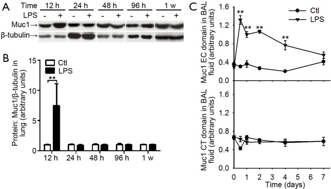
Figure 4.
Muc1 levels in lungs and BAL fluids from mice received intranasal LPS inhalation. (A) Expression of Muc1 protein relative to β-tubulin in lung treated with PBS (taken as normal control) or LPS inhalation was determined at indicated time by Western blotting with an antibody recognizing Muc1 CT domain. Representative blots for Muc1 and β-tubulin; (B) mean protein expression for Muc1 relative to β-tubulin. Values are presented as mean ± SEM and analyzed with Student’s t-test. **P<0.01 for LPS treated sample vs. respective control (n=5 mice in each group); (C) the levels of Muc1 CT and EC domain in BAL fluid at indicated time points were measured by ELISA. Values are presented as mean ± SEM and analyzed with Student’s t-test. **P<0.01 (n=5 mice per group). Muc1, mucin 1; BAL, bronchoalveolar lavage; LPS, lipopolysaccharide; CT, cytoplasmic tail; EC, extracellular.

