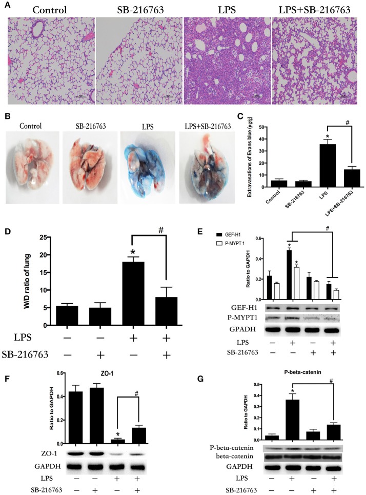
Figure 7.
Inhibition of GSK-3beta ameliorates LPS-induced ALI and vascular barrier dysfunction in mice. C57BL/6J mice were challenged with vehicle or LPS (2 mg/kg, i/t) for 24 h with or without SB-216763 pretreatment (20 mg/kg, i/p) (2 h prior to LPS intratracheal instillation) histological analysis of lung tissue by hematoxilin & eosin staining (× 100 magnification) (A). Evans blue dye (30 ml/kg, i/v) was injected 2 h before termination of the experiment (B). The quantitative analysis of Evans blue extravasation was performed by spectrophotometric analysis of Evans blue extracted from the lung tissue samples (C). *P < 0.05 vs. negative control. #P < 0.05 vs. corresponding LPS-stimulated group. Wet/dry ratio of lungs from the control, SB-216763 group, LPS group and LPS + SB-216763 group was represented as a histogram according to data (D). Expression of GEF-H1 and P-MYPT 1 in lung tissue samples was evaluated by Western blot (E). Expression of ZO-1 and P-beta-catenin in lung tissue samples was evaluated by Western blot analysis (F,G). *P < 0.05 vs. negative control. #P < 0.05 vs. corresponding LPS-stimulated group.

