Fig. 1.

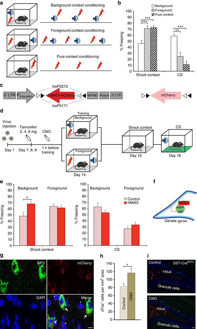
Pharmacogenetic inactivation of HIPP cells increases background context conditioning. a Schematic of the context conditioning paradigms. b Contextual and cued fear memory of C57Bl/6 mice trained according to those protocols. The contextual fear response is significantly lower in the background context group (n = 8) than in both foreground context (n = 8) and pure context (n = 8) groups in agreement with previous reports 4, 5, 9. At the same time, freezing to the CS is higher in the background context group, as expected. c Conditional hM4Di-mCherry and mCherry-control vectors. d Schematic of viral transduction and behavioral testing paradigms, depicting the virus injection, the activation of CRE recombination with increasing doses of tamoxifen and the activation of hM4Di with clozapine-N-oxide (CNO). Background and foreground context conditioning were applied to different groups and contextual and auditory memory was tested in both 24 h and 48 h later, respectively. e (Left) Memory to the background context is increased when HIPP cells are silenced during background context conditioning in SST-CreERT2 mice (n = 8; controls n = 10), whereas no effect is observed upon HIPP cells silencing during foreground conditioning (n = 8; controls n = 7). (Right) As expected the CS response is high in the background context group compared to the foreground context group. Viral intervention has no effect on cued memory, as the response to auditory stimuli remains unaltered in both training paradigms. f A schematic of the proposed circuitry. g Representative microscopic images show the adenoviral expression of mCherry-tagged hM4Di receptors in NPY+ cells (arrows) and NPY− cells of the hilus (asterisks). For an overview of the viral expression please see Supplementary Fig. 1d. Scale bar, 10 μm. h The number of cFos+ granule cells is increased after background context conditioning if hM4Di targeted HIPP cells are silenced with CNO (n = 6 each). i Representative images depicting cFos labeling and mCherry-tagged hM4Di in the DG following background context conditioning. Scale bar, 50 μm. Data are means + s.e.m. Statistical analysis was done with Fisher’s LSD following one-way ANOVA in a and with Student’s unpaired t-test in e, h. *P < 0.05; **P < 0.01; ***P< 0.001
