
Figure 1.
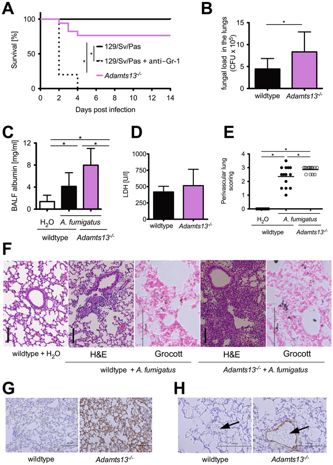
A. fumigatus infection of Adamts13 −/− mice results in increased mortality and fungal burden of the lungs. Wildtype (129\Sv\Pas) and Adamts13 −/− mice were infected i.t. with A. fumigatus (107 conidia per animal). (A) Overall survival were monitored for 14 days. The cumulative results of three independent experiments (129/Sv/Pas, n = 15; 129/Sv/Pas + Gr-1, n = 10; Adamts13 −/−, n = 17) are depicted. (*) indicates a significant difference (p < 0.05) by Mantel-Cox-Test. (B) 24 h after infection, some mice were sacrificed and their lungs were prepared and homogenized. The fungal burden was determined by plating serial dilutions of lung homogenates on Sabouraud 4% glucose agar plates. After 48 h the resulting colony-forming units (CFU) were enumerated. Shown are the cumulative results of three independent experiments (129/Sv/Pas, n = 10; Adamts13 −/−, n = 11) plus SD. (*) indicates a significant difference (p < 0.05) by Mann-Whitney U-test. (C) The albumin concentration of the BAL fluid was quantified by ELISA 24 h after infection. The cumulative results of two (H2O-treated 129/Sv/Pas, n = 6), three (A. fumigatus-treated Adamts13−/−, n = 11) or four independent experiments (A. fumigatus-treated 129/Sv/Pas, n = 13) plus SD are depicted. (D) Plasma LDH activity was measured 24 h after infection of wildtype (n = 4) and Adamts13 −/− (n = 5) mice. (E–H) 24 h after infection, three mice of the indicated groups were sacrificed, paraffin sections of the lungs were prepared and stained with H&E, Grocott (F), C3d antibody (G) and VWF antibody (H). (E) Perivascular inflammation were scored by S. R. who was blinded to the experimental treatment groups. (*) indicates a significant difference (p < 0.05) by one-way ANOVA with Bonferroni’s posttest. (F–H) Representative images of lung sections are depicted (H&E, Grocott and C3d 400x magnification, VWF 800x magnification), vessels are marked by an arrow.