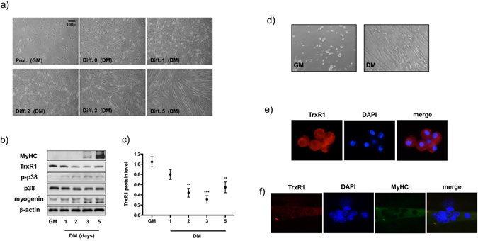
Figure 1.

TrxR1 is downregulated during skeletal muscle differentiation. (a) Representative image of C2C12 myoblasts differentiation process. Growth medium (GM) is maintained in proliferation conditions (Prol) until 90% confluency. Pictures were taken respectively at 0, 1, 2, 3 and 5 days after switching to differentiation media (DM). (b) Western blot analysis showing the TrxR1 protein contents in C2C12 cells cultivated in GM and DM (1, 2, 3, 5 days) conditions. Antibodies against Myogenin, p38, p-p38 and MyHC were used to monitor myogenesis process, whereas β-actin was employed as a housekeeping loading control. (c) Relative quantification of TrxR1 protein expression normalized to β-actin levels of western blots in figure b. The TrxR1/β-actin ratio in GM conditions is set = 1. Results are represented as mean ± SEM of three independent experiments. (d) Images (10x magnification) of murine satellite cells in proliferation (GM) and 5 days after switching to differentiation media (DM). (e) Representative image (63x magnification) of the immunofluorescence analysis showing TrxR1 expression in satellite cells and in (f) myotubes originated from them. Multinucleated myotubes are positive for MyHC and DAPI staining was used to detect nuclei.
