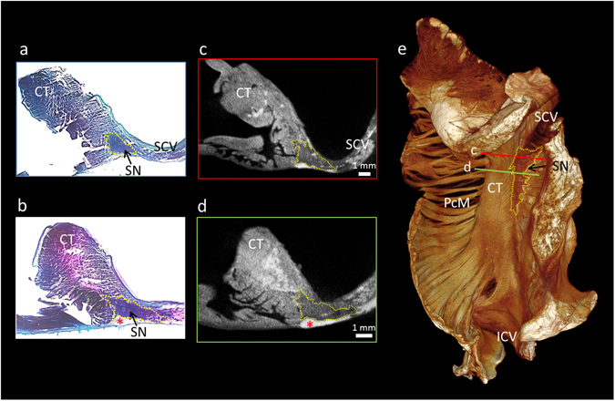
Figure 1.

Micro-CT allows objective discrimination of the human sinus node. This figure demonstrates high resolution (28 × 28 × 28 µm3) micro-CT data from part of the right atrium containing the sinus node. For this figure, the sinus node is outlined in yellow in short-axis micro-CT sections (c,d) and in matching histological sections taken from the same sample (a,b). The plane of section in c and d is shown on the 3D volume rendering (endocardial view) (e). The sections are available to view without the outlines in the supplementary Fig. S2. CT- terminal crest, ICV- inferior caval vein, PcM- pectinate muscles, SCV- superior caval vein, SN- sinus node, *- indicates epicardial fat.
