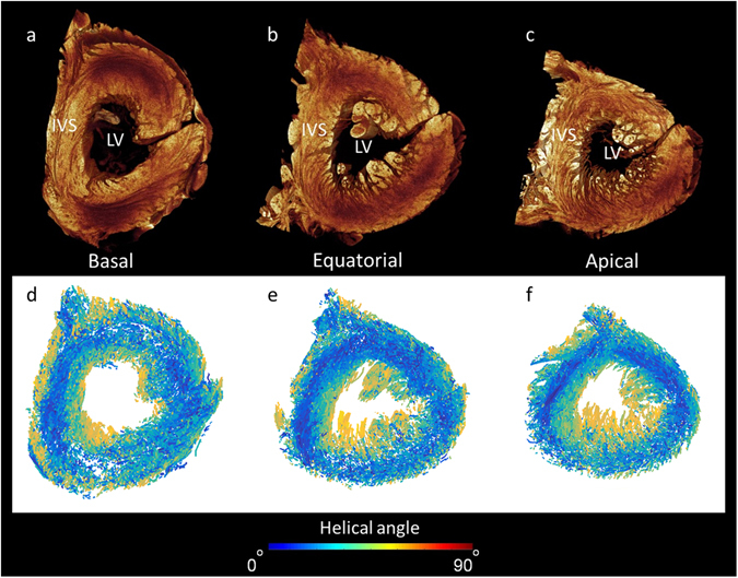
Figure 8.

Extraction of cardiomyocyte orientation in the human left ventricle. Panels a–c show volume renderings in short-axis view taking from the basal, equatorial, and apical regions. Panels d–f show corresponding cardiomyocyte orientation in which the absolute helical angles derived from the CT dataset are coded in colour (see colour map). Note the right ventricular free wall is removed from view. IVS- interventricular septum, LV- left ventricular cavity.
