Figure 4.

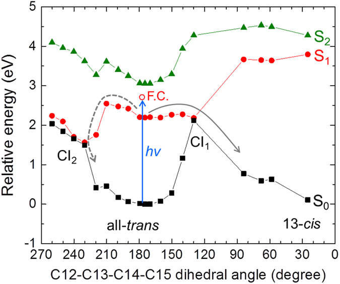
CASPT2//CASSCF(12,12) photoisomerization pathways from the all-trans retinal conformer, when the retinal Schiff base is hydrogen-bonded to Glu162. S0 (black closed squires with black solid line), S1 (red closed dots with red solid line) and S2 (green closed triangles with green solid line) energy profiles are shown. The blue arrow shows light excitation of all-trans retinal as performed on our pump-probe and pump-dump-probe spectroscopic experiments. The open red dot shows the Franck–Condon state (F.C.) The solid gray arrow shows the isomerization path through energy barrier of 1.96 kcal/mol (0.09 eV) on S1 via a conical intersection CI1. The dashed gray arrow indicates a relaxation path through energy barrier of 7.42 kcal/mol (0.32 eV) on S1 via another conical intersection CI2.
