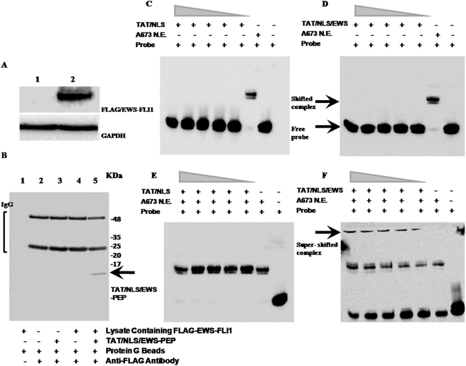
Figure 7.

Peptide-protein and Peptide-Protein-DNA interactions of the TAT/NLS/EWS-PEP peptide. (A) Western blot analysis of ectopically expressed FLAG/EWS-FLI1 expression in MCF-7 cell lysates. Lanes 1: lysate from vector alone expressing cells, and 2: Lysate from FLAG/EWS-FLI1 expressing cells. Upper panel blot was probed with anti-FLAG mouse monoclonal antibody, and the lower panel shows the same blot reprobed with mouse monoclonal anti-GAPDH antibody. (B) Detecting TAT/NLS/EWS-PEP in FLAG immunoprecipitates (IP) using western blot: Lanes 1–4: 1: Bead control, 2: IP from MCF-7 empty vector expressing cells, 3: IP from MCF-7 empty vector expressing cells in the presence of TAT/NLS/EWS-PEP, and 4: IP from MCF-7 FLAG/EWS-FLI1 expressing cells and 5: IP from MCF-7 FLAG/EWS-FLI1 expressing cells in the presence of TAT/NLS/EWS-PEP. Full length blots are displayed in the figure. (C) EWS-FLI1 EMSA using 7XGGAA duplex probe. TAT/NLS peptide interaction with 7XGGAA probe. Lane 1(from right): probe alone, Lane 2: probe and A673 N.E., Lanes 3 to 7: probe with TAT/NLS at increasing concentrations (1, 2, 4, 8 and 10 μM). (D) EWS-FLI1 EMSA using 7XGGAA duplex probe. TAT/NLS/EWS-PEP peptide interaction with 7XGGAA probe. Lanes 1: probe alone, 2: probe and A673 N.E., Lanes 3 to 7 probe with TAT/NLS/EWS-PEP at increasing concentrations (1, 2, 4, 8 and 10 μM). (E) EWS-FLI1 EMSA using 7XGGAA duplex probe. TAT/NLS peptide interaction with 7xGGAA probe in presence of A673 N.E. Lane 1: probe alone, Lane 2: probe and A673 N.E, Lanes 3 to 7 mixture of probe with A673 N.E. and TAT/NLS at increasing concentrations (1, 2, 4, 8 and 10 μM). (F) EWS-FLI1 EMSA using 7XGGAA duplex probe. TAT/NLS/EWS-PEP peptide interaction with 7XGGAA probe in presence of A673 N.E., Lane 1: probe alone, Lane 2: probe and A673 N.E., Lane 3 to 7 mixture of probe with A673 N.E., andTAT/NLS/EWS-PEP at increasing concentrations (1, 2, 4, 8 and 10 μM).
