Fig. 4.
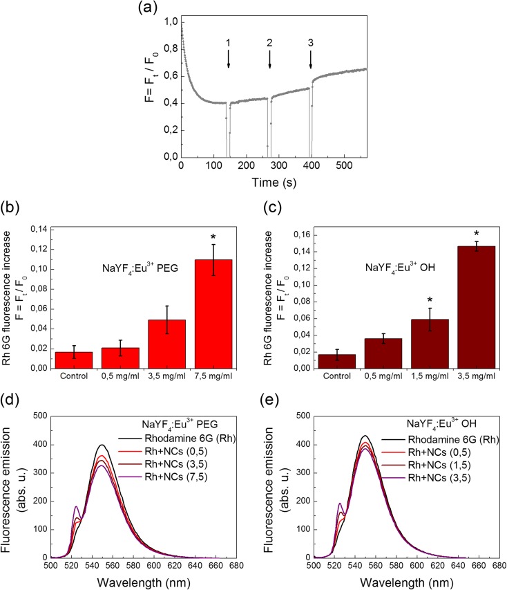
The membrane potential of the nerve terminals after the addition of NaYF4:Eu3+-PEG (a). The suspension of the synaptosomes was equilibrated with potential-sensitive dye rhodamine 6G (0.5 μM); when the steady level of the dye fluorescence had been reached, NaYF4:Eu3+-PEG at a concentrations of 0.5, 3.5 and 7.5 mg/ml in three additions (arrow nos. 1, 2 and 3) were applied to the synaptosomes. Trace represents three experiments performed with different preparations. An increase in the fluorescence signal of rhodamine 6G in response to application of (b) NaYF4:Eu3+-PEG (0.5–7.5 mg/ml) or (c) NaYF4:Eu3+-OH (0.5–3.5 mg/ml), respectively, to the synaptosomes. Data is mean ± SEM. Single asterisk indicates P < 0.05 as compared to control (baseline fluorescence). Fluorescence emission spectra of rhodamine 6G (0.5 μM) in the standard salt solution before and after application of (d) NaYF4:Eu3+-PEG (0.5–7.5 mg/ml) or (e) NaYF4:Eu3+-OH (0.5–3.5 mg/ml)

