Figure 1.
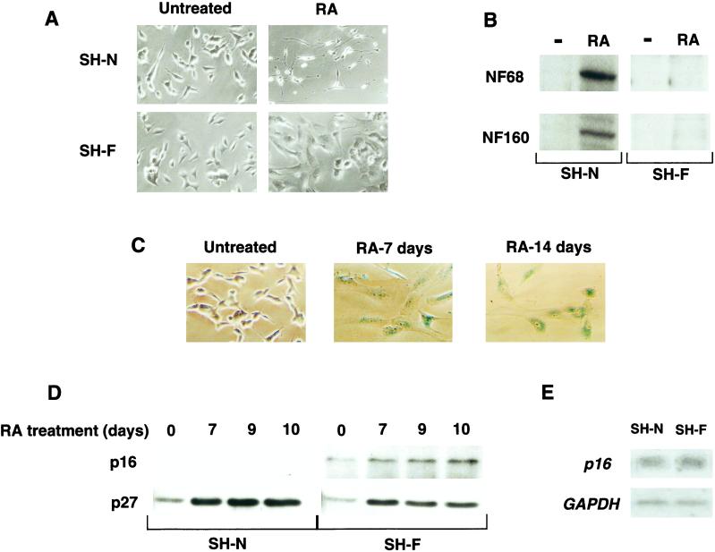
Effects of RA on the NB cell subclones SH-N and SH-F. (A) Cellular morphology of SH-N and SH-F cells left untreated or treated with RA for 7 days. (B) Neurofilament 68 and neurofilament 160 Western immunoblot analysis of SH-N and SH-F left untreated (−) or treated with RA for 7 days (RA). (C) SH-F cells were treated with RA for the indicated times and stained with 5-bromo-4-chloro-3-indol β-d-galactopyranoside to measure SA-β-gal activity. (D) p16INK4a and p27Kip1 Western immunoblot analysis of SH-N and SH-F treated with RA for the indicated times. (E) Northern blot analysis for p16INK4a and glyceraldehyde-3-phosphate dehydrogenase (GAPDH) of untreated SH-N and SH-F.

