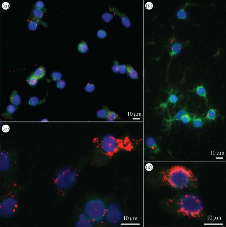
Figure 3.
Visualization of Wolbachia in A. vulgare haemocytes by FisH. Wolbachia are labelled in red, cytoskeleton in green (Phalloidin) and nucleus in blue (DAPI). Pictures illustrate the strong increase in infection intensity in haemocytes between (a) P1, (b) P2 and (c,d) P3. (Online version in colour.)

