Figure 4.
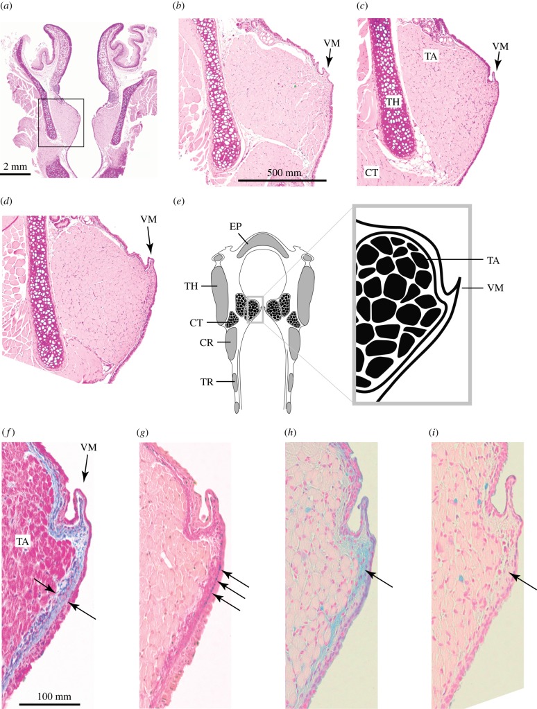
Grasshopper mouse larynx reveals vocal membranes. (a) Coronal cross section of a larynx. (b–d) Higher magnification images (from the square in (a) of three male mice vocal fold cross sections (H&E stain). On the medial edge of both vocal folds, all mice had vocal membranes consisting of a double layer of epithelium with a small amount of connective tissue between the epithelial layers. (e) Schematic of a grasshopper mouse larynx. (f,g) Lamina propria of vocal membranes contains collagen (blue stain in f), elastin (black stain in g) and hyaluronan (blue stain in h). (i) Removal of hyaluronan by hyaluronidase digestion with subsequent AB staining. CR, cricoid cartilage; CT, cricothyroid muscle; EP, epiglottis; TA, thyroarytenoid muscle; TH, thyroid cartilage; TR, tracheal rings; VM, vocal membrane. (Online version in colour.)

