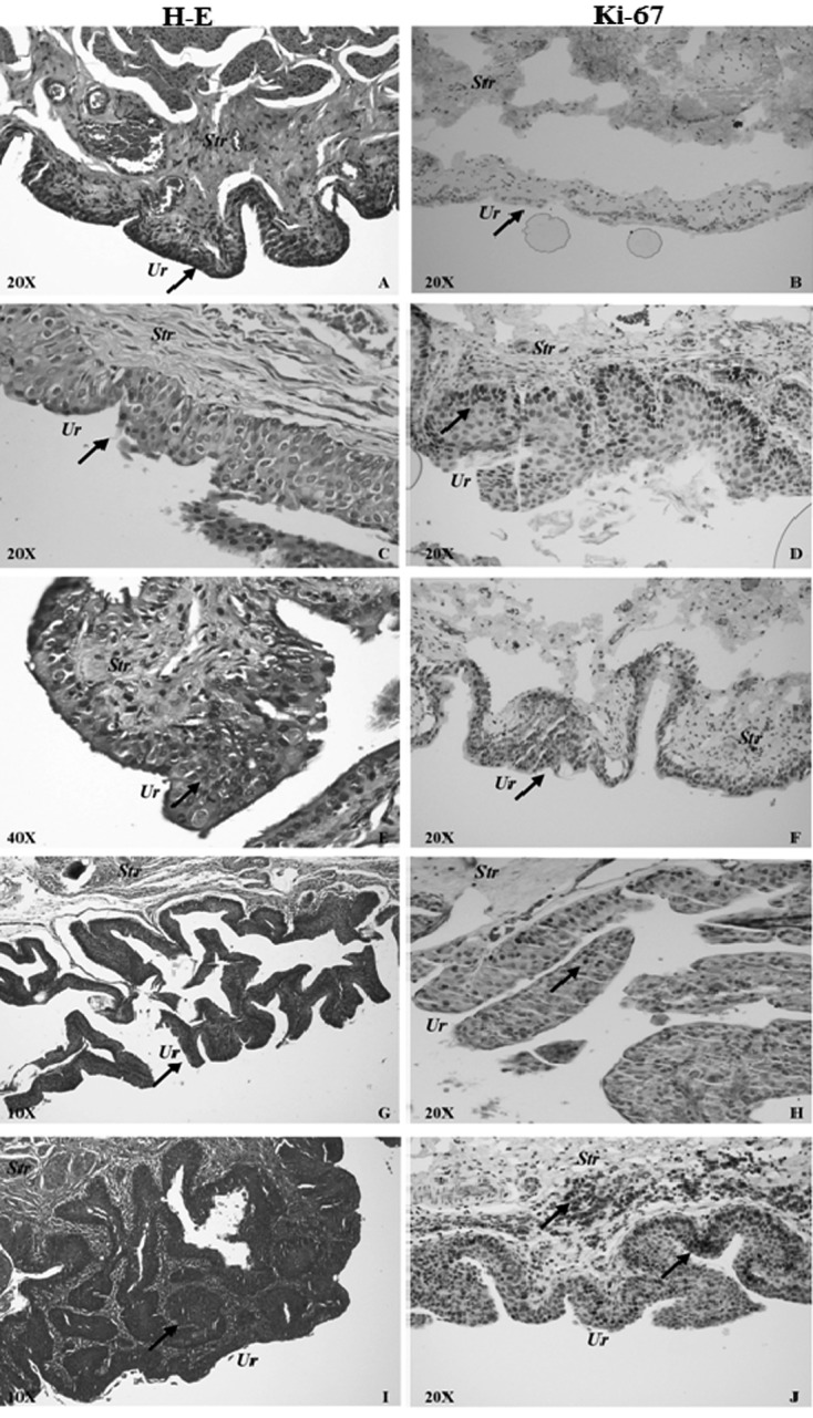
Fig. 2.

Normal urothelium and representative pictures of different stages of MNU-induced male rat bladder carcinogenesis in the MNU-only and MNU+O3 groups with H-E stain (left column) and Ki-67 immunolabeling (right column). A, B) Normal urothelium from the MNU+O3 group. Arrows show a normal urothelium. C, D) Dysplastic urothelium from the MNU+O3 group. Arrows show a dysplastic urothelium in cells stained with H-E (C) and Ki-67 (D). E, F) High-grade intraepithelial neoplasia (CIS) from the MNU-only group. Arrows show the CIS area (E) and CIS cells stained with Ki-67 (F). G, H) Low-grade pTa papillary urothelial carcinoma from the MNU+O3 group. Arrows show the pTa carcinoma area (G) and Ki-67 staining of it (H). I, J) High-grade pT1 papillary urothelial carcinoma from the MNU-only group. Arrows show the pT1 carcinoma area (I) and corresponding Ki-67 staining in the urothelium and submucosa (J). Str, Stroma; Ur, Urothelium.
