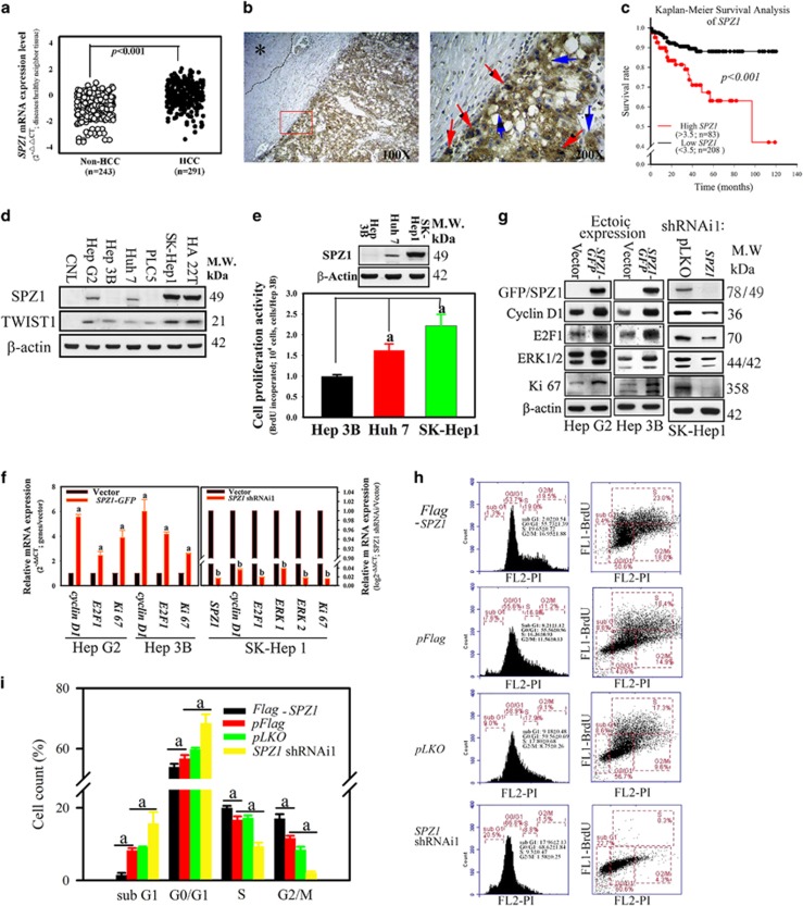
Figure 1.
Correlation of SPZ1 with cell proliferation and poor prognosis in HCC tumor patients. (a) Expression of SPZ1 mRNA was compared between HCC samples (n=291) and non-HCC samples (n=243). (b) Expression of SPZ1 protein in HCC samples. Brown, SPZ1 expression; asterisk, area of healthy hepatocytes; blue arrows, SPZ1 expression in cytoplasm; red arrows, SPZ1 expression in nuclei. (c) Kaplan–Meier survival analysis of SPZ1. Red, patients with higher expression (n=83; >3.5-fold than the control region) of SPZ1; black, patients with lower expression (n=208; <3.5-fold than the control region) of SPZ1. (d) Endogenous expression of SPZ1 and TWIST1 in various hepatoma cells. (e) SK-Hep1 with high SPZ1 expression showing greater proliferative activity. (f) Effect of forced and knockdown expression of SPZ1 on cyclin D1, E2F1, Ki67 and ERK1/2 mRNAs. (g) Effect of ectopic and knockdown expression of SPZ1 on cyclin D1, E2F1, Ki67 and ERK1/2 proteins. (h) Serum-starved Hep G2 transformed cells (5 × 10⁵ cells) with pFlag-SPZ1, pFlag, pLKO and SPZ1 shRNAi1 were cultured in DMEM plus 10% FBS for 24 h, stained with BrdU-specific antibody and propidium iodide (PI) and subjected to Fluorescence-Activated Cell Sorting (FACS) analysis to determine the percentage of cells in each phase of the cell cycle. FL1-BrdU, bromodeoxyuridine stained cells; FL2-PI, PI stained cells. (i) Percentages of cells that were stained with BrdU-specific antibodies. The percentage of each cell cycle was measured. Means±s.d. of results of three experiments are shown. a and b, P<0.001.

