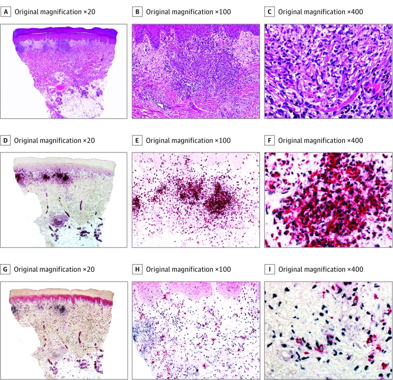
Figure 2. Histopathologic and Immunohistochemical Features of Histiocytoid Sweet Syndrome (Case 8).
A-C, Hematoxylin-eosin–stained specimens. A, Scanning power shows edema and nodular infiltrates in the superficial dermis. B, The infiltrate is close to the epidermis, but there is no exocytosis. C, The nodules are mostly composed of mononuclear cells with twisted vesicular nuclei and scant eosinophilic cytoplasm. D-F, Double immunostained specimens (myeloid nuclear differentiation antigen [MNDA, black] and myeloperoxidase [MPO, red]). D, Scanning power. E, With this double immunostain, exocytosis of single cells is more evident than with hematoxylin-eosin. F, Most of the cells of the infiltrate coexpress MNDA in their nuclei and MPO in their cytoplasm. G-I, Double immunostained specimens (MNDA [black] and CD163 [red]). G, Scanning power. H, With this double immunostain, most of the cells express MNDA in their nuclei, while less scattered cells express CD163 in their cytoplasm. I, Higher magnification demonstrates that cells expressing CD163 in their cytoplasm do not express MNDA in their nuclei.

