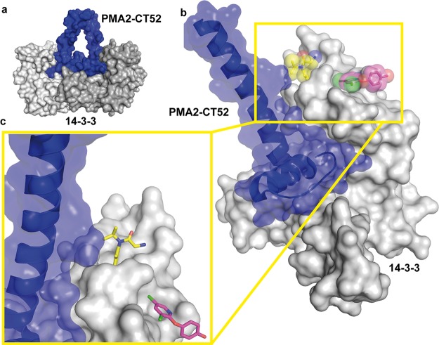
Figure 7.
Position of the secondary binding sites of NV1 and NV2 in relation to the tobacco 14-3-3/PMA2-CT52 complex. (a) Overview of the complex of the tobacco T14-3c dimer (gray surface) bound to two PMA-CT52 molecules (blue surface) (PDB entry 2O98). (b) Superimposition of the 14-3-3 structure with NV1 (yellow spheres and sticks) and NV2 (magenta spheres and sticks), with PMA2-CT52 (blue cartoon and semitransparent surface) bound to one monomer of T14-3c (gray surface). (c) Detailed view of the main helix of CT52 binding in the vicinity of the NV1 pocket in 14-3-3.

