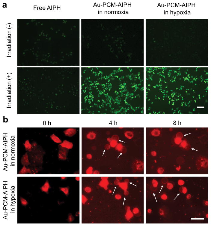
Figure 3.
a) Detection of AIPH-induced ROS in A549 cells by DCFHDA staining. The A549 cells were treated with AIPH (20 μg·mL−1) or Au-PCM-AIPH (AIPH: 20 μg·mL−1) in normoxic and hypoxic culture media, respectively, for 2 h and then irradiated by an 808-nm laser for 30 min. Scale bar = 50 μm. b) Morphology of A549 cells after incubation with Au-PCM-AIPH (AIPH: 20 μg·mL−1) for 2 h and irradiated with an 808-nm laser for 30 min, and then incubated for another 4 h and 8 h in normoxic and hypoxic media, respectively. The cells were stained with a lipophilic membrane dye Dil to visualize the morphological change. Scale bar = 50 μm.

