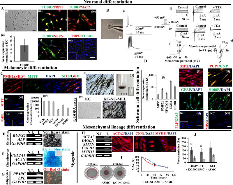
Figure 3. KC-NC in vitro differentiate into functional neural crest derivatives.
KC-NC differentiate into functional peripheral neurons (A). Phase image of KC-NC derived neurons (A,i). KC-NC neurons expressed the TUBB3 gene (A,ii) and positively immunostained for PRPH (Peripherin) and TUBB3 (A,iii) suggestive of their peripheral origin. KC-NC neurons expressed MAP2 (A,iv) and NEUN (A,v). KC were negative for neuronal markers (A,vi; IgG control is shown in inset). KC-NC neurons are electrophysiologically mature (B). KC-NC neurons displayed evoked action potentials upon 50pA step current injections (n=15 cells) (B,i,ii). KC-NC neurons expressed tetrodotoxin (TTX) sensitive inward Na+ currents (n=4 cells) (B,iii) and tetraethylammonium (TEA) sensitive outward K+ currents (n=10 cells) (B,iv) . Red lines under traces in (B,iii) and (B,iv) represent portions of traces expanded for visual ease. Current-voltage relationship for K+ currents (B,v) and Na+ currents (B,vi) are shown (error bars, SEM). KC-NC differentiate into functional melanocytes (C). MITF and PMEL (HMB45) dual positive melanocytes (C,i), melanin containing melanocytes (C,ii) and centrifuged melanocyte pellet (C,iii). KC-NC derived melanocytes upregulated key melanocyte markers (TYR, PMEL, DCT, MITF, GPNMB, S100B) (C,iv) and produced melanin in response to L-DOPA (C,v). KC lacked melanocyte markers. (Cvi,vii; IgG control is shown in inset). KC-NC differentiate into Schwann cells (D) as shown by increased expression of Schwann cell specific genes (MPZ, PMP22, NFATC4, EGR2, S100B) (D,i) and positive immunofluorescence for MPZ (D,ii), CNPase and PLP1 (D,iii), GFAP (D,iv), S100B (D,v), . KC were negative for Schwann cell markers (Dvi,vii; IgG control is shown in inset). KC-NC differentiate into functional mesenchymal lineages i.e. osteocytes (E), chondrocytes (F) and adipocytes (G) as shown by RT-PCR and functional stains for each lineage. KC-NC give rise to functional smooth muscle cells as confirmed by gene and protein expression for SMC markers (H). Compaction of fibrin hydrogels embedded with KC-NC-SMC. The area of the hydrogels was imaged and quantified using image J. Aortic smooth muscle cells were used as positive control (I). Fibrin-based tissue constructs fabricated with KC-NC-SMC displayed isometric contractions in response to vasoagonists (U46619, Endothelin-1 and KCl). ASMC were used as positive control (J). Scale bars, 50µm. All values are mean±SD. $p < 0.05. Experiments were repeated three times independently. Inset images are uninduced controls. N.I. = non-induced; I. = induced

