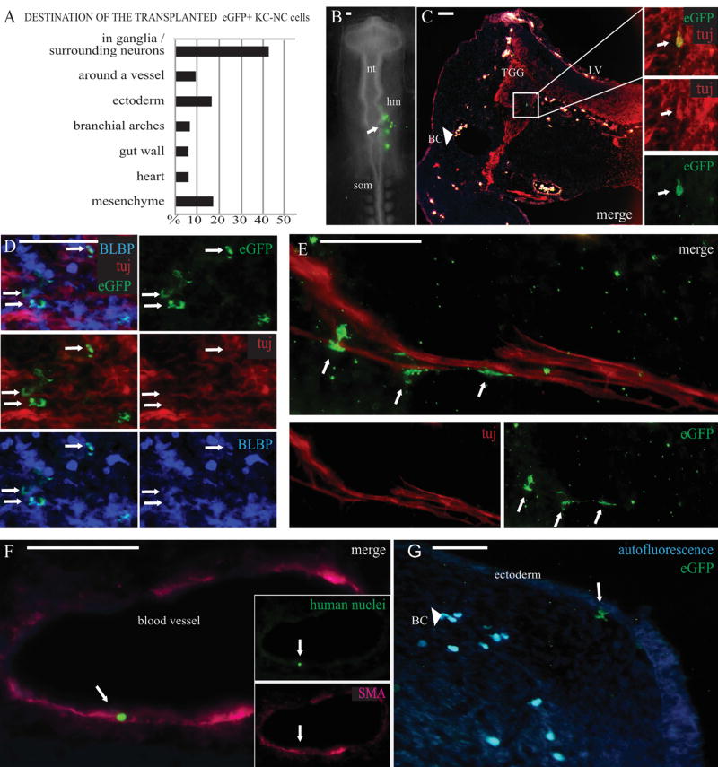
Figure 5. KC-NC migrate and differentiate into neural crest lineages in ovo.
KC-NC migrate and differentiate into neural crest lineages in ovo as shown in 12µm transverse sections of 2-3 days old chicken embryos. Percentages of transplanted cells detected in each target structure in the developing chick embryos (n=8 embryos; total number of detected EGFP+ cells = 151 out of ~ 400 transplanted cells) (A). The EGFP+ KC-NC or KC were transplanted to the head mesenchyme (hm) of 10-13 somite host chick embryos on Day 0 and the cells were traced 2-3 days post transplantation (B). A Tuj/EGFP double positive neuron in the trigeminal ganglion (TGG) (day 3). The transplanted EGFP+ cells are not visible on other channels and thus are not to be mixed with highly autofluorescent blood cells (BC) that are found throughout the mesenchyme in vessels and small capillaries; merged image with red, blue and green fluorescence makes the blood cells look white (C). BLBP+/EGFP+ double positive glial cells in the TGG (day 3) (D). EGFP+ putative Schwann cells localized around a nerve bundle at the outer edge of a cranial ganglion (day 3) (E). A cranial blood vessel surrounded by αSMA+ cells with one of them originating from the transplanted cells as indicated by co-expression of the human specific nuclear marker (day 2) (F). Differentiating EGFP+ putative melanocytes were detected under the cranial ectoderm (day 3). Blood cells are highly autofluorescent on both green and blue channels (light blue) (G). Scale bar 50 µm. hm= head mesenchyme, nt=neural tube, som= somites, LV=lateral ventricle, TGG=trigeminal ganglion, BC=blood cells.

