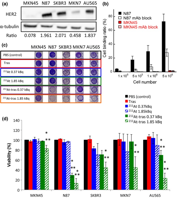
Figure 1.

In vitro analysis of cell binding by astatine‐211‐labeled trastuzumab (211At‐tras) and subsequent cell death. (a) Human epidermal growth factor receptor 2 (HER2) expression in MKN45, N87, SKBR3, MKN7, and AU565 cells. α‐Tubulin was used as a loading control. Ratios of the band intensity of HER2 relative to α‐tubulin are indicated below the images. (b) Cell binding ratio of 211At‐trastuzumab to N87 and MKN45 cells with/without trastuzumab (mAb) block. Bars are labeled in the graph. Three independent experiments were carried out, each in triplicate (n = 3). (c) Representative images of stained surviving cells at 7 days after 24 h of treatment with PBS control, unlabeled trastuzumab (Tras), 211At (0.37 or 1.85 kBq), or 211At‐tras (0.37 or 1.85 kBq). (d) Quantification of the viability of the cells in panel (c) treated with 211At‐tras for 7 days. Bars are labeled in the graph. Three independent experiments were carried out in triplicate (n = 3). All data represent mean ± SD. *P < 0.05 versus control; **P < 0.05 versus Tras.
