
Figure 6.
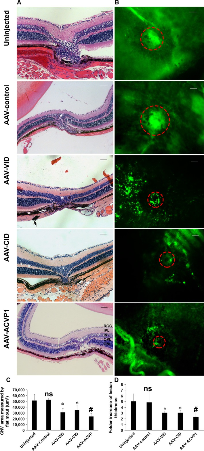
Assessment of choroidal neovascularization (CNV) area by H&E staining and choroid/RPE flatmount of laser‐induced CNV mice. (A) Representative images of H&E staining taken by Zeiss microscope. ONL, outer nuclear layer; OPL, outer plexiform layer; INL, inner nuclear layer; IPL, inner plexiform layer; RGC, retinal ganglion cell layer. Original magnification 20× Bar = 50 μm. The laser lesions were marked by star, and the choroid neovascularization were indicated by dashed circles. (B) Representative choroid/RPE flatmount images were taken by Zeiss fluorescence microscope, and (C) area were measured by Zeiss AxioVision Software measurement tool. Original magnification 20× Bar = 100 μm. Values on y‐axis represent CNV area (μm2). (D) Statistical analysis of neovascular lesion thickness from each group. Ratio of lesion thickness to adjacent normal thickness is measured. Values on y‐axis represent fold increase in lesion thickness. Data expressed as mean ± SD; (n = 5 per group); ns means not significant versus uninjected group *P < 0.05 (versus uninjected group). #P < 0.05 (versus AAV‐VID and AAV‐CID group).