Figure 7.

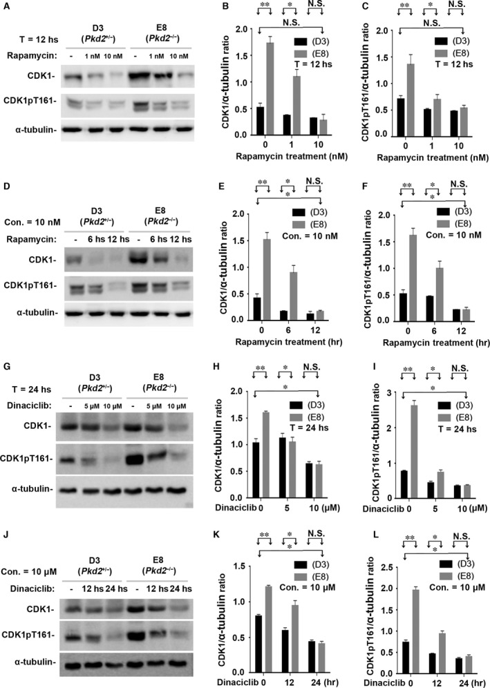
Rapamycin suppresses CDK1 and its activated form CDK1pT161 in vitro. (A) The same cell lines (E8 and D3) were also incubated with 0, 1, or 10 nM rapamycin for 12 hrs. Western blots of the lysates of E8 and D3 cells showed that rapamycin decreased the expression of CDK1 and its activated form CDK1pT161 in a dose‐dependent manner. (B–C) Normalized quantitative analysis using the densitometry values from the Western blots in (A). (D) The cell lines were also incubated with 10 nM rapamycin for 0, 6 or 12 hrs. Western blots of the cell lysates showed that rapamycin significantly decreased the expression of CDK1 and its activated forms CDK1pT161 in a manner dependent on the duration of incubation. (E–F) Normalized quantitative analysis using the densitometry values from the Western blots presented in (D). Other cell‐cycle‐associated CDKs (such as CDK2, CDK4 and CDK6) were not affected by PC2 expressional levels and rapamycin treatment (data not shown). (G) The same cell lines were cultured with CDK inhibitor Dinaciclib with 0, 5 and 10 μM for 24 hrs. Western blots of the cell lysates showed that Dinaciclib significantly inhibited the expression of CDK1 and its activated forms CDK1pT161 in a dose‐dependent manner. (H–I) Normalized quantitative analysis using the densitometry values from the Western blots presented in (G). (J) The cell lines were also cultured with CDK inhibitor Dinaciclib with 10 μM for 0, 12 or 24 hrs. Similar results to different concentrations were seen in Western blot assays on the different durations of Dinaciclib incubation. (K–L) Normalized quantitative analysis using the densitometry values from the Western blots presented in (J). N.S. = No significance; *P < 0.05; **P < 0.01.
