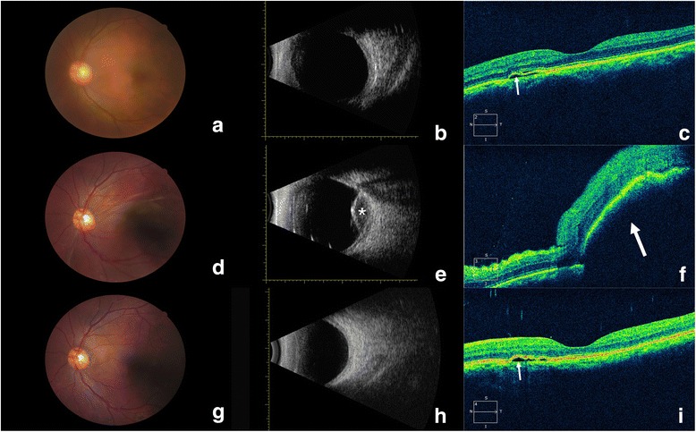
Fig. 2.

Changes in the eye with uveal effusion. a, b Preoperative fundus photography (a) and type B ultrasonography (b) were unremarkable. c Preoperative OCT revealed fluid accumulation (arrow) beneath the neurosensory retina, resulting in mild serous detachment. d Fundus photography on postoperative day 1 revealed suprachoroidal exudation in the macula. e Type B ultrasonography on postoperative day 1 revealed serous choroidal detachment, with little blood at the posterior pole (asterisk). f OCT on postoperative day 1 revealed a bulging macular choroid with edematous neuroepithelium and subfoveal effusion (arrow). (g, h) Fundus photography (g) and type B ultrasonography (h) on postoperative day 50 were unremarkable. i OCT on postoperative day 50 revealed complete resolution of the uveal effusion and mild serous detachment of CSC (arrow)
