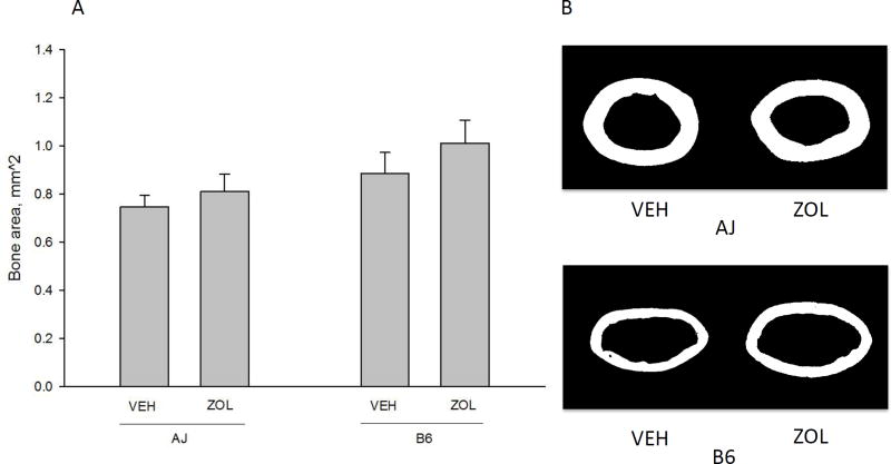
Figure 2.
Cortical bone area of the femoral diaphysis. There was a significant main effect of genotype (B6 had higher bone area compared to A/J, p = 0.0001) and drug (zoledronate (ZOL) led to significantly higher bone area compared to vehicle (VEH), p = .0001). There was no significant interaction between variables (p = 0.076). Representative images (from the animal within each group closest to the group mean) of diaphyseal bone for each genotype/drug condition are shown for comparison. All data presented as means and standard deviations.

