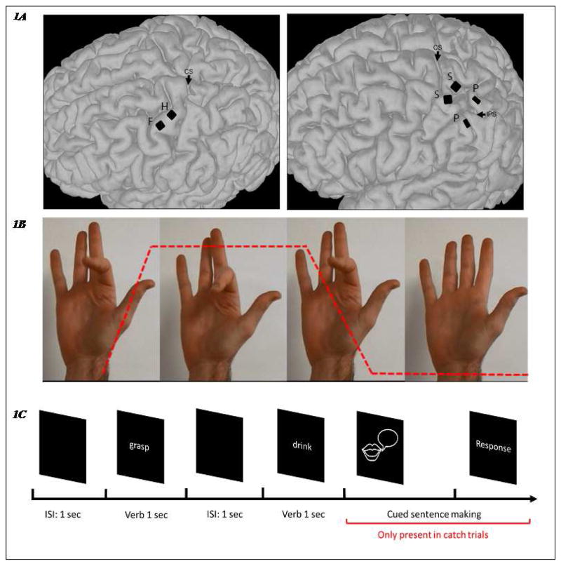
Figure 1.
The microelectrode array locations and the experimental paradigm. 1A: Array locations of the two subjects, registered on their respective MRI scans. Left: Subject A, one array was implanted in the finger area of the primary motor cortex (precentral gyrus, labeled ‘F’) and another was in the hand area (labeled ‘H’). Both arrays have 96 recording channels. CS: central sulcus. Right: Subject B, two square arrays (96 recording channels) were implanted in the primary somatosensory area (postcentral gyrus, labeled ‘S’) while another two rectangle arrays (32 recording channels) were placed on superior and inferior parietal lobules, respectively (labeled ‘P’). IPS: intraparietal sulcus. 1B: Experimental Paradigm for the video-following task: videos with different hand, wrist, elbow and shoulder movements were shown to the subjects to map sensorimotor cortex responses on the same day as participants completed the verb-reading task. These videos were made following the same timeline: each video consisted of one second of movement and two seconds of holding. Each video consisted of five such repetitions. An example is shown here. The red dashed line indicates the kinematics of index finger movement. Both subjects were instructed to watch all of the videos and to ‘attempt’ the movements in the video as if they could perform them, at the same pace as the video. 1C: Experimental Paradigm for the verb-reading task: the subjects were instructed to read verbs silently. The verbs were presented for 1 second with an inter-stimulus interval of 1 second. Catch trials were included to keep the subjects attentive. In a catch trial, they were cued to make a sentence with the verb just shown.

