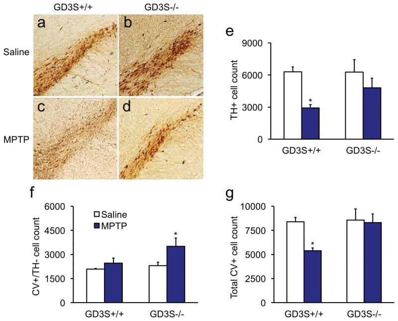
Figure 4. Tyrosine hydroxylase expression is partially suppressed in GD3S−/− mice.
a,c,e) Wild-type mice lost 53.0 ± 4.8% of the TH+ neurons in the SNc after the third MPTP injection regimen. b,d,e) GD3S knockouts had a 22.5 ± 14.6% reduction of TH+ neurons, which was not statistically significant. f) Interestingly, GD3S−/− mice lesioned with MPTP had more CV-positive neurons in the SNc that were negative for TH. g) The total CV+ neurons (positive or negative for TH) did not differ in GD3S−/− mice compared to their saline-treated controls, suggesting that TH expression was suppressed in surviving SNc neurons. Data are expressed as mean ± SEM. *p < .05.

