
Figure 3.
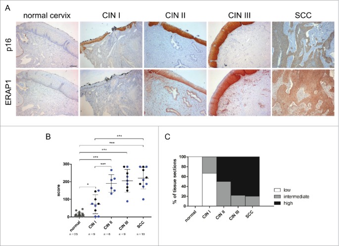
Immunohistochemical analysis of ERAP1 expression in cervical tissue. A, Human tissue sections were stained for ERAP1 and p16, a surrogate marker for HPV oncogene expression. Protein expression was analyzed in histologically normal cervical epithelium (normal cervix), in HPV16-positive dysplastic CIN (cervical intraepithelial neoplasia) I to CIN III lesions and in SCC (cervical squamous cell carcinoma). Scale bar corresponds to 200µm. B, Immunohistochemical scores were determined by multiplying the frequency score of ERAP1 expressing cells with the maximum intensity score of ERAP1 expression. Each symbol represents one sample, symbols in gray represent HPV-negative samples, symbols in blue represent HPV16-transformed tissue, and symbols in black represent tissue transformed with other HPV high-risk types. Means ± SD are indicated. *p ≤ 0.05, ***p ≤ 0.001 (ANOVA multiple comparison test). C, Immunohistological scores were equally categorized into low (0–95), intermediate (96–190) and high expression (191–285).